坤灵最新logo-scaled-1.png)
Introduction
Microtomes are essential precision instruments in pathology laboratories, enabling the preparation of ultra-thin tissue sections for microscopic examination. From routine histopathology to advanced research applications, microtomes play a vital role in ensuring diagnostic accuracy.
This guide explores working principles, classifications, applications, and microtome maintenance best practices, helping laboratories improve performance, compliance, and efficiency.
I. Definition and Overview of Microtomes
A microtome is a precision cutting instrument used to produce thin tissue sections (typically 1–50 μm) for microscopic analysis.
Key Terminology
- Microtome: Tissue sectioning instrument
- Sectioning: Cutting thin tissue slices
- Paraffin Embedding: Wax infiltration for support
- Cryosectioning: Frozen tissue cutting
- Ribbon: Continuous strip of tissue sections
II. Working Principle of Microtomes
Microtomes operate through controlled mechanical advancement + blade cutting:
- Sample Advancement: 1–50 μm per step
- Cutting Motion: Rotary / sliding / rocking
- Section Collection: Transfer to glass slides
- Temperature Control (Cryostat): -15°C to -30°C
III. Primary Functions in Pathology
- Diagnostic tissue preparation
- IHC and special staining
- Frozen section analysis
- Research and 3D reconstruction
- Biobanking and archival studies
IV. Structural Features and Materials
- Heavy-duty base (anti-vibration)
- Precision feed system
- Adjustable blade holder
- Safety lock & blade guard
- Ergonomic handwheel design
V. Classification and Technical Parameters
| Type | Thickness (μm) | Application | Power | Weight |
|---|---|---|---|---|
| Rotary | 1–60 | Routine histology | Manual / Motor | 25–40 kg |
| Cryostat | 5–50 | Frozen sections | 500–800W | 150–250 kg |
| Sliding | 1–100 | Large tissues | Manual | 30–50 kg |
| Ultramicrotome | 0.05–1 | Electron microscopy | 100–150W | 50–70 kg |
VI. Application Industries
- Hospitals & pathology labs
- Pharmaceutical companies
- Research institutes
- Veterinary labs
- Forensic laboratories
VII. Installation & Compliance
- Temperature: 18–22°C
- Humidity: 40–60% RH
- ISO 14644 cleanroom compliance
- GMP validation (IQ/OQ/PQ)
- IEC 61010 safety standards
VIII. Microtome Maintenance
Proper microtome maintenance is critical for ensuring section quality, instrument longevity, and regulatory compliance.
1. Daily Maintenance
- Clean debris after each use
- Wipe surfaces with 70% ethanol
- Remove paraffin residues
- Check blade condition
2. Weekly Maintenance
- Inspect blade holder alignment
- Clean feed mechanism
- Lubricate moving components (if required)
3. Monthly Maintenance
- Verify section thickness accuracy
- Inspect mechanical stability
- Check handwheel locking system
4. Annual Maintenance
- Full calibration by certified technician
- Replace worn components
- Verify safety systems
5. Cryostat-Specific Maintenance
- Defrost regularly
- Replace HEPA filters (every 3–6 months)
- Clean internal chamber to prevent contamination
Microtome Maintenance Best Practices
- Use only manufacturer-approved consumables
- Keep detailed maintenance logs (GMP requirement)
- Train operators for proper handling
- Schedule preventive maintenance
👉 Key Insight:
Proper maintenance can reduce downtime by 30–40% and significantly improve section consistency.
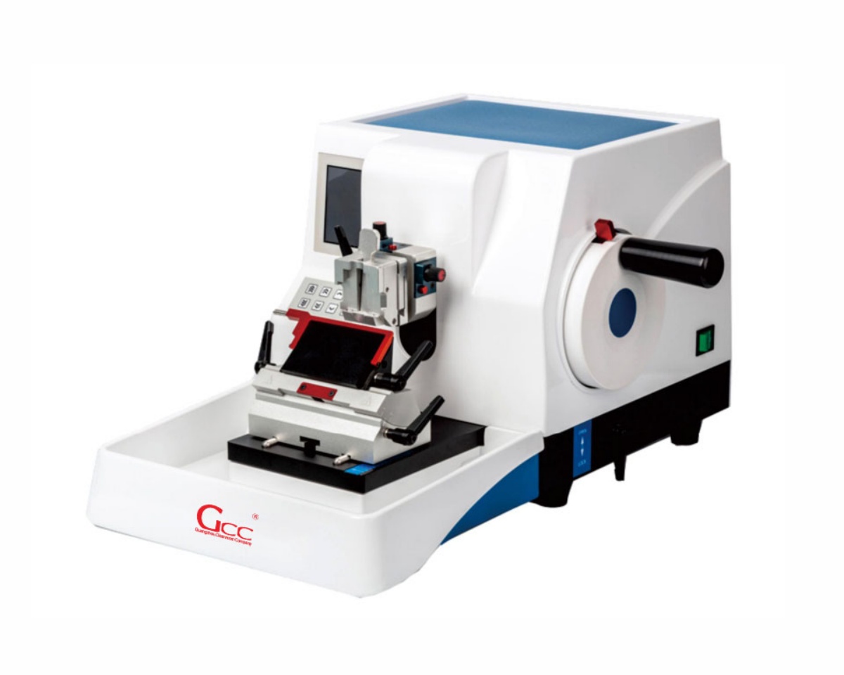
IX. GCC Microtome – Real Equipment Showcase
To better understand real-world performance, here is a GCC pathology microtome in operation:
High-Resolution Equipment Images
GCC Microtome Key Advantages
- High-precision sectioning (±1 μm accuracy)
- Stable mechanical structure (low vibration)
- Ergonomic design for long operation
- GMP-ready documentation support
- Compatible with modern histopathology workflows
X. Conclusion
Microtomes are indispensable tools in modern pathology, enabling precise and reliable tissue sectioning.
By combining:
- Proper equipment selection
- Standardized operation
- Structured maintenance programs
Laboratories can achieve higher diagnostic accuracy, improved efficiency, and full regulatory compliance.
Enhance Your Pathology Laboratory with GCC Solutions
GCC Pathology provides:
- Advanced microtomes
- Histopathology workflow solutions
- Cleanroom-compatible lab equipment
- Full GMP validation support
👉 Contact us today to receive a customized pathology solution tailored to your laboratory.