坤灵最新logo-scaled-1.png)
Introduction
In histopathology laboratories, tissue sectioning is a critical step before microscopic examination. Technicians must prepare tissue samples carefully to observe cellular structures under a microscope.
Before sectioning begins, laboratory staff process the tissue through fixation, dehydration, and paraffin embedding. These steps stabilize the sample and make it suitable for thin sectioning.
A precision instrument called a microtome performs this task. Understanding the microtome working principle helps technicians produce consistent and high-quality tissue sections. Section quality directly affects diagnostic accuracy and research results.
Modern microtomes have evolved significantly. Early models relied only on mechanical movement. Today’s systems combine precision mechanics with digital control technology.
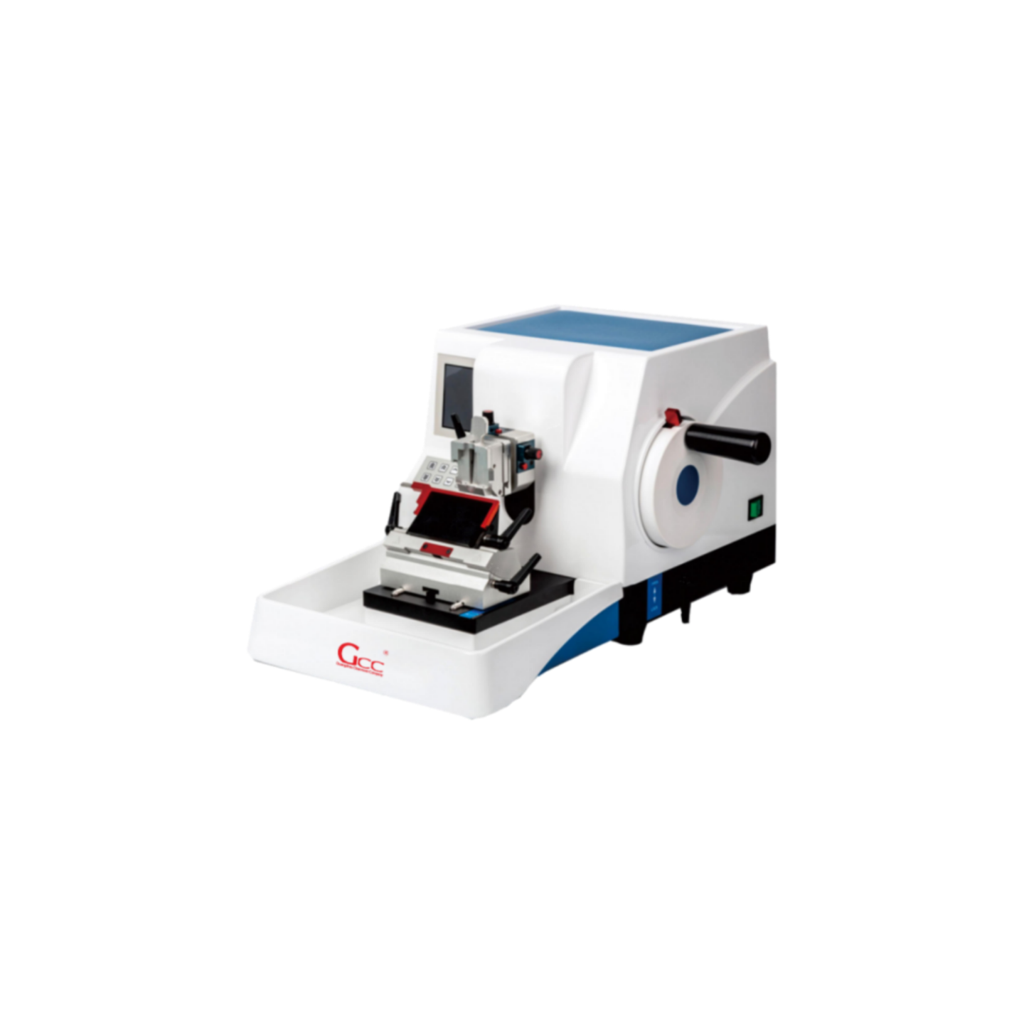
The GCC-QPJ-1200 Series Microtome & Cryostat offers reliable and accurate tissue sectioning for pathology laboratories, histology research centers, and clinical diagnostic laboratories.
Microtome Working Principle
The microtome working principle relies on precise mechanical feeding and cutting.
First, the technician fixes the prepared tissue block into the specimen holder. Most samples are embedded in paraffin to provide stability during cutting.
The operator then rotates the handwheel or activates the cutting system. This action moves the specimen holder toward the blade along a controlled path.
At the same time, the feeding mechanism advances the specimen by a very small distance. This distance determines the thickness of each tissue section.
As the specimen passes the blade, the blade cuts extremely thin layers from the tissue block. These thin slices form ribbon-like strips.
Technicians collect these ribbons and place them into a warm water bath. The warm water helps flatten the sections.
After that, technicians transfer the flattened sections onto glass slides. The slides then go through staining and microscopic examination.
This precise and continuous process allows laboratories to produce uniform and high-quality tissue sections for histopathological analysis.
Core Technology of the GCC-QPJ-1200 Series Microtome
The GCC-QPJ-1200 Series Microtome & Cryostat integrates advanced technologies to improve accuracy, efficiency, and usability.
The instrument features an HD color touch screen. The interface allows technicians to control the system easily and monitor operation data in real time.
Users can quickly switch between slicing mode and trimming mode. This function simplifies workflow and improves laboratory efficiency.
The system uses a high-precision feeding mechanism with a 25,600 fine fraction controller and photoelectric sensor. This design allows sectioning accuracy of up to 0.1 μm.
Technicians can also choose flexible feeding modes. The system supports continuous movement from 0.1 to 3.5 mm/s. It also supports single-step movement between 1 and 100 μm. These options provide better control during trimming and sectioning.
For safety, the instrument includes an independent 360° handwheel locking system. Operators can lock the handwheel at any position to prevent accidental movement.
The microtome also provides a slice counting function. This feature helps laboratory staff track section numbers and manage workflow records.
Despite its advanced technology, the system maintains a compact design. It saves valuable laboratory space while delivering stable performance.
Technical Specifications
The GCC-QPJ-1200C Microtome meets the operational needs of modern histopathology laboratories.
The instrument measures 580 × 490 × 315 mm. Its compact size allows easy installation on standard laboratory workbenches.
Technicians can adjust the trimming thickness from 0 to 999 μm with 1 μm increments. This range supports different sample preparation requirements.
The system provides specimen retraction from 0 to 100 μm. Users can adjust or disable this feature depending on the application.
The fixed section length ranges from 1 to 100 μm. This function ensures accurate control of section thickness.
The microtome supports specimens up to 50 × 60 × 40 mm. The vertical movement range of the specimen reaches 60 mm.
The blade holder allows left-right movement and front-back travel of 28 ±1 mm. This design helps technicians adjust blade positioning easily.
Technicians can adjust the section angle between 0° and 10° to optimize cutting performance.
Both coarse feeding speed and precision feeding speed range from 1 to 3.5 mm/s, which ensures smooth and stable sectioning.
Applications
The GCC-QPJ-1200 Series Microtome serves many laboratory environments that require high-precision tissue sectioning.
Typical applications include:
- Pathology laboratories
- Histology research centers
- Clinical diagnostic laboratories
- Medical research institutions
- Universities and teaching laboratories
These laboratories rely on microtomes to prepare thin tissue sections for microscopic observation, disease diagnosis, and biomedical research.
Conclusion
The microtome working principle uses a precise feeding and cutting mechanism to produce extremely thin tissue sections. Technicians gradually advance the specimen toward a sharp blade to achieve consistent thickness.
Modern microtomes combine mechanical precision with digital control systems. These improvements increase accuracy and simplify laboratory workflows.
The GCC-QPJ-1200 Series Microtome & Cryostat integrates advanced feeding technology, intelligent control, and user-friendly operation. These features help laboratories produce reliable tissue sections and maintain high diagnostic standards.
As a result, the GCC-QPJ-1200 has become an important instrument for pathology laboratories, research institutions, and clinical diagnostic facilities that require accurate and stable tissue sectioning.