坤灵最新logo-scaled-1.png)
In a hospital, there is a department that seems mysterious to patients but plays a crucial role in clinical care—the Pathology Department. At the heart of this department lies the pathology technical laboratory, which serves as its core production workshop. Unlike the operating room, which is thrilling, or the imaging department, which provides intuitive visual results, the work here forms the “gold standard” for final diagnoses. Consequently, it guides treatment decisions as the department’s “strategic command center.”
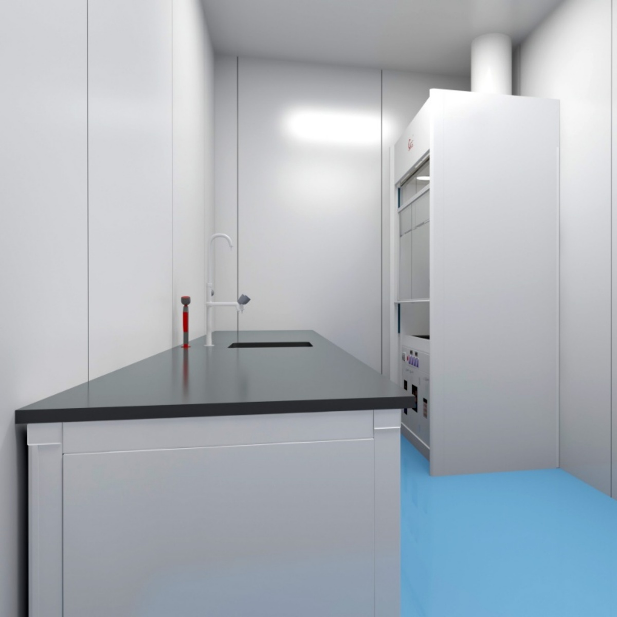
If we compare the final pathology report to an authoritative “forensic certificate,” the pathology technical laboratory actively processes and presents all the “evidence.” From an engineering perspective, it functions as a highly specialized, automated, and standardized “biological tissue precision processing factory.”
I. Core Mission: From Tissue to High-Quality Glass Slide
The laboratory’s primary task transforms human tissue samples, obtained via surgery or biopsy, ranging from a few millimeters to several centimeters, into transparent glass slides only 4–5 microns thick (about 1/20th the diameter of a human hair). Each slide must meet these engineering standards:
- Structural Integrity: The cells, tissue layers, and spatial relationships remain intact.
- High Transparency: The slide allows clear observation under a microscope.
- Accurate Staining: Different tissue components appear distinctly.
Only when these criteria are satisfied can pathologists make precise diagnoses under the microscope.
II. Production Line: A Rigorous Standardized Process
The slide-making process follows a tightly controlled assembly line, and any error in a single step may ruin the “product” and delay diagnosis.
1. Specimen Receipt and Fixation (Raw Material Intake & Preservation)
- Operation: Technicians first verify specimen information and perform preliminary handling. They then immerse the tissue in formalin fixative, which is the most critical step.
- Principle: Fixation acts as “biological preservation.” By cross-linking proteins, it halts all cellular biochemical activity immediately, preserving the tissue’s state at the moment of collection and preventing decay. Therefore, this step lays the foundation for all subsequent processes.
2. Dehydration, Clearing, and Wax Infiltration (Pre-Embedding Processing)
- Operation: Technicians place fixed tissues in a series of alcohol solutions with increasing concentration for dehydration. Next, they immerse the tissue in clearing agents like xylene to replace the alcohol and finally infiltrate the tissue with molten paraffin.
- Principle: Tissue contains water, which is incompatible with paraffin. This chemical exchange replaces water molecules with paraffin, hardening the tissue and enclosing it in wax, which prepares it for sectioning. In other words, it is like replacing the water in a sponge with molten plastic; once cooled, the sponge gains shape and rigidity.
3. Embedding (Creating Standardized Blocks)
- Operation: Wax-infiltrated tissue blocks are placed into molds filled with liquid wax, oriented precisely, and cooled to solidify.
- Principle: This process produces uniform, adequately hardened blocks, which serve as standard “workpieces” for microtomy. As a result, the sections remain consistent and operable.
4. Sectioning and Mounting (Ultra-Precision Cutting and Transfer)
- Operation: Technicians mount the blocks on a microtome and slice them into thin ribbons. They gently flatten the ribbons using a brush and a water bath, then transfer them onto slides.
- Principle: Microtomes perform micron-level precision cutting. Technicians carefully control the water bath temperature within ±1°C. If the temperature is too low, ribbons wrinkle; if it is too high, tissue may burn. Therefore, this step resembles the “nano-level lathe machining” of biology.
5. Staining and Coverslipping (Adding Contrast and Permanent Preservation)
- Operation: Technicians apply Hematoxylin & Eosin (HE) staining. Slides are sequentially stained with hematoxylin (blue nuclei) and eosin (pink cytoplasm/collagen), dehydrated, cleared, and mounted with coverslips.
- Principle: Staining adds a “color filter” that highlights tissue components, making cells and structures easily distinguishable under the microscope. Coverslipping protects the slide for long-term observation.
III. Advanced “Industrial Equipment” and Technology
Modern pathology laboratories rely heavily on automation, moving beyond purely manual operations:
- Fully Automated Tissue Processors: These machines precisely control time, temperature, and solution changes, standardizing dehydration and replacing labor-intensive manual work.
- Automated Stainers: Technicians pre-program staining workflows, ensuring consistency and eliminating human error.
- Immunohistochemistry (IHC) Instruments: These devices label specific proteins (e.g., HER2, Ki-67, PD-L1) for cancer subtyping, prognosis assessment, and targeted therapy guidance. Therefore, IHC forms the engineering foundation for precision medicine.
IV. Quality Control: The Lifeline of Engineering
Pathology laboratories strictly follow ISO15190 and other quality management standards. Technicians verify specimens, monitor reagents, and perform batch slide QC. In essence, they act as both operators and quality engineers, ensuring every slide meets high standards of accuracy and reliability.
Conclusion
The pathology technical laboratory is a hidden “precision factory” behind every diagnosis, integrating mechanical engineering, chemical engineering, materials science, and biology. Each pathologist technician actively transforms cold biological tissue into vivid, information-rich slides. Although they rarely meet patients directly, they serve as indispensable engineers, illuminating the path toward accurate treatment and safeguarding patient health.