坤灵最新logo-scaled-1.png)
In histopathology, diagnostic accuracy relies heavily on the quality of tissue sections. As a cornerstone of the pathology laboratory, a microtome directly influences whether sections meet the standards required for molecular-level observation.
In this guide, we explain the working principle of microtomes, explore application scenarios for different models, and outline best practices to improve laboratory efficiency.
1. What is a Pathology Microtome?
A pathology microtome is a high-precision instrument that cuts biological tissues embedded in paraffin or other media into ultra-thin, transparent sections (typically 3–5 micrometers).
Technicians then mount these sections on glass slides and stain them for microscopic examination. As a result, pathologists can accurately evaluate cellular structures and detect abnormalities.
2. How It Works: The Microtome Principle
Modern microtomes rely on precise mechanical movement to produce consistent sections. More specifically, the cutting process involves three key steps:
- Specimen Advancement
The system advances the specimen toward the blade with each cycle according to the preset thickness. This ensures consistent section thickness. - Tangential Sectioning
The blade moves vertically (or the specimen moves against it), creating a clean slicing action through controlled shear force. - Consistency Assurance
A stable structure prevents vibration and uneven feeding. Therefore, the microtome produces uniform sections without “skipping,” which is essential for downstream Immunohistochemistry (IHC).
3. Primary Types and Application Scenarios
Laboratories choose different microtomes based on specific diagnostic needs. Below are the most common types:
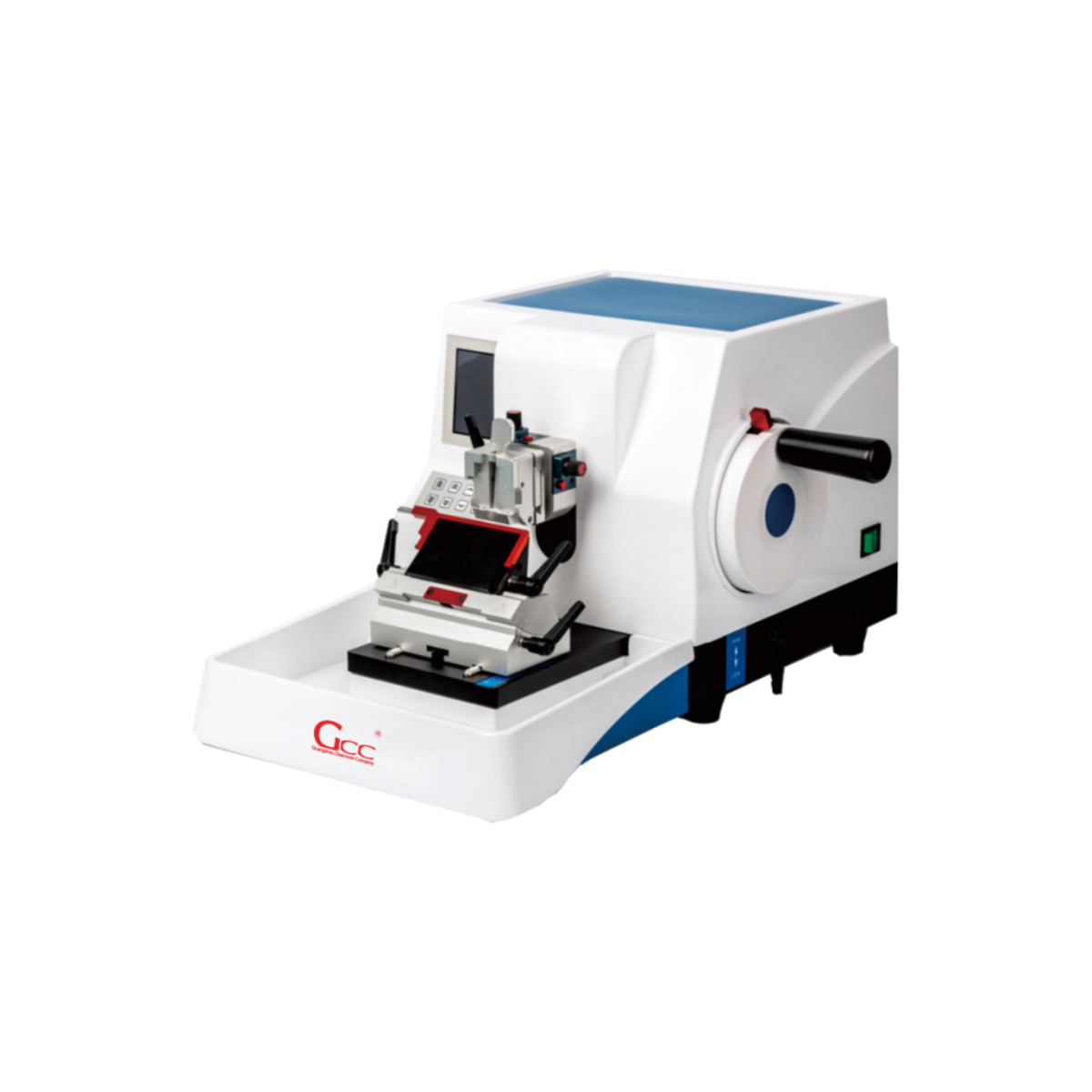
Rotary Microtome
This type dominates pathology laboratories.
- Core Use: Routine paraffin-embedded tissue sectioning
- Advantages:
- Enables smooth and continuous ribboning
- Handles high workloads efficiently
- Provides consistent section thickness
In addition, a heavy and stable base reduces vibration and improves cutting precision.
Cryostat Microtome
This system supports rapid diagnosis.
- Core Use: Intraoperative (frozen section) diagnosis
- Advantages:
- Eliminates dehydration and embedding steps
- Delivers results within 15–20 minutes
- Preserves tissue morphology in a frozen state
Therefore, surgeons can make immediate clinical decisions during procedures.
Sliding Microtome
This type handles specialized applications.
- Core Use: Large specimens or hard tissues (e.g., woody samples)
- Advantages:
- Accommodates oversized blocks
- Provides stable cutting for dense materials
4. Best Practices for Superior Section Quality
Even with advanced equipment, technicians must follow proper techniques to achieve optimal results. For example:
- Blade Selection
Choose blades based on tissue hardness. At the same time, inspect and replace blades regularly to maintain sharpness. - Specimen Cooling
Cool paraffin blocks on an ice plate before sectioning. Consequently, the tissue gains better support and cuts more cleanly. - Angle Adjustment
Set the clearance angle between 5° and 8°. This adjustment improves cutting efficiency and prevents compression or chatter.
5. GCC: Precision Solutions for Pathology Laboratories
At GCC, we recognize that every section affects diagnostic outcomes. Therefore, we design our microtomes to deliver consistent, high-precision performance under demanding laboratory conditions.
Our systems feature:
- Corrosion-resistant materials for long-term durability
- High-precision transmission mechanisms for stable operation
- Robust structural design to minimize vibration
Moreover, we provide integrated laboratory solutions tailored to your workflow. Whether you are building a new lab or upgrading existing equipment, our team ensures seamless integration and reliable performance.
📞 Contact Our Technical Specialists
If you are planning a new pathology lab or upgrading your microtome system, our experts are ready to help.
- WhatsApp: +86 18148635992
- Email: Sales@gzkunling.com
- Website: www.gccpathology.com