坤灵最新logo-scaled-1.png)
The pathology laboratory is one of the core diagnostic departments in a hospital. A scientifically designed and properly zoned laboratory is essential for workflow efficiency, diagnostic quality, and biosafety. Below is a detailed overview of the main functional areas in a pathology laboratory, including their roles and operational connections.
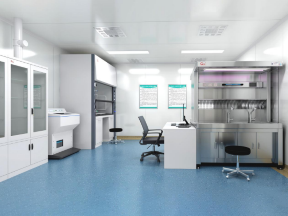
1. Specimen Receiving and Registration Area
Primary Functions:
- Receive pathology specimens from surgical, outpatient, and inpatient departments
- Verify patient information, specimen details, and requisition forms
- Assign unique specimen identification numbers and register them
- Conduct preliminary assessment of specimen integrity and fixation
Operational Connection:
After registration, specimens are sent to either the routine grossing room or the rapid frozen section area according to type and urgency.
2. Specimen Storage Room (Specimen Repository)
Primary Functions:
- Short-term storage of received specimens awaiting processing
- Provide proper storage conditions, such as refrigeration
- Maintain specimen quality during storage
Operational Connection:
Specimens are delivered from storage to the corresponding grossing area according to the processing schedule.
3. Routine Grossing Room
Primary Functions:
- Systematically dissect and examine routine pathology specimens
- Select representative tissue blocks for embedding
- Record detailed gross morphological features
- Photograph special specimens for documentation
Operational Connection:
Processed tissue blocks are sent to the tissue processing area for dehydration and paraffin embedding.
4. Frozen Section Grossing Area
Primary Functions:
- Handle intraoperative rapid frozen specimens
- Quickly select diagnostic tissue blocks
- Support rapid intraoperative diagnosis through frozen section techniques
Operational Connection:
Selected tissue blocks are immediately transferred to the frozen section room for rapid slide preparation.
5. Tissue Processing Room
Primary Functions:
- Perform dehydration, clearing, and paraffin infiltration using automated tissue processors
- Ensure tissue reaches suitable hardness for sectioning
- Standardize processing to maintain slide quality
Operational Connection:
Processed tissue blocks and embedding cassettes are sent to the embedding room.
6. Embedding Room
Primary Functions:
- Place paraffin-infiltrated tissue into molds and embed in paraffin blocks
- Ensure proper tissue orientation
- Label paraffin blocks accurately with specimen information
Operational Connection:
Finished paraffin blocks are sent to the sectioning room for microtomy.
7. Sectioning Room
Primary Functions:
- Cut paraffin blocks into uniform thin sections (typically 3–5 µm)
- Float sections on warm water and mount onto slides
- Dry slides to secure tissue sections
Operational Connection:
Prepared slides are sent to the staining room.
8. Staining Room
Primary Functions:
- Perform routine hematoxylin-eosin (H&E) staining
- Complete staining manually or using automated stainers
- Ensure consistent and stable staining quality
Operational Connection:
Stained slides are sent to the coverslipping room.
9. Coverslipping Room
Primary Functions:
- Apply neutral mounting medium and cover slips to stained sections
- Ensure proper mounting without bubbles
- Guarantee appropriate mounting medium volume
Operational Connection:
Coverslipped slides are sent to the pathologist reading area.
10. Special Staining Room
Primary Functions:
- Perform special stains such as reticulin, Masson, PAS, etc.
- Aid pathologists in differential diagnosis
- Highlight specific tissue components or pathogens
Operational Connection:
Specially stained slides are sent directly to the pathologist reading area.
11. Immunohistochemistry (IHC) Room
Primary Functions:
- Detect specific protein expression using antigen-antibody reactions
- Assist in tumor classification, prognosis evaluation, and therapeutic target identification
- Ensure reproducible results through standardized procedures
Operational Connection:
IHC-stained slides are sent to the pathologist reading area.
12. Molecular Pathology Laboratory
Primary Functions:
- Perform molecular testing such as gene analysis and FISH
- Provide molecular-level diagnostic information for precision medicine
- Strictly separate areas to prevent cross-contamination (reagent preparation, sample handling, amplification, product analysis)
Operational Connection:
Molecular test results are integrated into the final pathology report.
13. Cytopathology Room
Primary Functions:
- Handle cytology specimens from body fluids, aspirates, etc.
- Prepare conventional or liquid-based cytology slides
- Perform cytological staining and preliminary screening
Operational Connection:
Cytology slides are sent to cytopathologists for evaluation.
14. Pathologist Reading Area
Primary Functions:
- Pathologists examine slides under a microscope and render diagnoses
- Conduct consultations for difficult cases
- Serve as a training area in teaching hospitals
Operational Connection:
Diagnostic results are sent to the report writing area.
15. Pathology Reporting Room
Primary Functions:
- Compile pathology reports based on diagnostic results
- Review reports for accuracy and completeness
- Issue and archive reports
Operational Connection:
Final reports are issued to clinical departments and archived.
16. Pathology Archives
Primary Functions:
- Long-term storage of paraffin blocks, slides, and reports
- Maintain a retrievable system for efficient access
- Control temperature and humidity for preservation
Operational Connection:
Receive and archive specimens and documents from all laboratory sections according to regulations.
17. Instrument and Equipment Room
Primary Functions:
- Store spare instruments and equipment
- Conduct routine maintenance and calibration
- Repair and service small laboratory devices
18. Reagent and Consumables Storage Room
Primary Functions:
- Store reagents, dyes, and consumables in an organized manner
- Separate storage by requirements (room temperature, refrigerated, hazardous)
- Monitor inventory and expiration dates
19. Office and Study Area
Primary Functions:
- Provide workspace for pathology staff
- Facilitate case discussions and professional learning
- Serve as consultation space for clinicians
20. Auxiliary Areas
Primary Functions:
- Locker rooms and showers for staff hygiene
- Temporary storage for medical waste
- Cleaning and disinfection area for instruments and containers
Functional Flow Summary
- Specimen Flow: Clinical departments → Specimen receiving → Specimen storage → Grossing → Tissue processing → Embedding → Sectioning → Staining → Coverslipping → Pathologist reading
- Information Flow: Requisition → Registration → Grossing record → Diagnostic notes → Pathology report → Clinical department
- Rapid Workflow: Intraoperative specimens → Frozen section grossing → Frozen section → Rapid staining → Intraoperative diagnosis → Operating room
- Auxiliary Workflow: Special specimens → Special staining/IHC/molecular testing → Supplementary diagnosis → Integrated into final report
Design Principles and Considerations
- Unidirectional Workflow: Prevent cross-contamination between clean and contaminated areas
- Biosafety Compliance: Separate clean, semi-contaminated, and contaminated zones
- Efficiency First: Position adjacent functional areas to minimize specimen transport distance
- Safety Measures: Store and manage chemical reagents and hazardous substances separately
- Future-Proofing: Reserve space for new technologies and procedures
A well-designed pathology laboratory optimizes workflow, improves diagnostic efficiency, ensures staff safety, and provides a solid foundation for high-quality pathology services. While hospital scale may affect functional area configuration, the core workflow and zoning principles should remain consistent.
Frequently Asked Questions
How to minimize formaldehyde exposure in pathology labs?
Minimize formaldehyde exposure through proper ventilation, formalin filling systems, down-draft exhaust on grossing stations, personal protective equipment, and regular air quality monitoring. Formaldehyde Exposure Guide
What ISO and quality standards apply to pathology laboratories?
Pathology labs typically comply with ISO 15189 (medical laboratories), CAP (College of American Pathologists) requirements, and GMP standards for quality management and operational consistency. Pathology Lab Quality Control
What biosafety level is required for a pathology lab?
Most clinical pathology labs operate at BSL-2 level, requiring biosafety cabinets for specimen handling, proper waste management, and adherence to standard precautions for infection control. Lab Safety Standards
What is the difference between ducted and ductless fume hoods?
Ducted fume hoods exhaust contaminated air outside the building through ductwork, ideal for high-volume chemical use. Ductless fume hoods use HEPA and activated carbon filtration to clean and recirculate air, offering flexible installation. Pathology Fume Hood Guide
What are the key standards for pathology laboratory design?
Pathology lab design must comply with ISO 15189, CAP, and local biosafety regulations. Key considerations include workflow zoning, pressure differentials, airflow management, and equipment layout. Pathology Lab Design Standards