坤灵最新logo-scaled-1.png)
Establishing an efficient and compliant pathology department requires a profound understanding of the diagnostic workflow. From the moment a tissue specimen enters the laboratory to the final microscopic diagnosis, every step relies on high-precision pathology laboratory instruments.
This guide outlines a comprehensive histology lab equipment list covering the entire process from grossing to staining, helping you optimize your laboratory setup for maximum accuracy and safety.
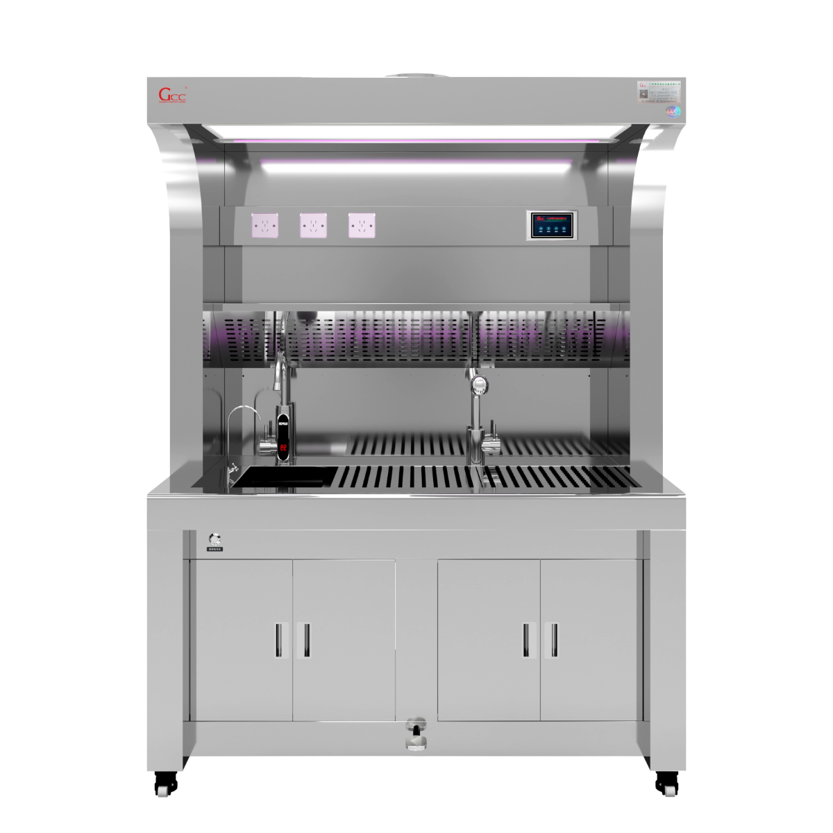
Phase 1: Fixation and Grossing
This is the starting point of pathological analysis. To ensure specimen integrity and protect personnel from hazardous formaldehyde vapors, high-performance ventilation is non-negotiable.
Pathology Grossing Station: As the heart of the lab, it must feature superior extraction capabilities (such as downdraft and backdraft ventilation). GCC’s Grossing Station Series is constructed from 304 stainless steel, integrating ergonomic design with automated flushing systems.
Learn More: Explore GCC Professional Grossing Station Product
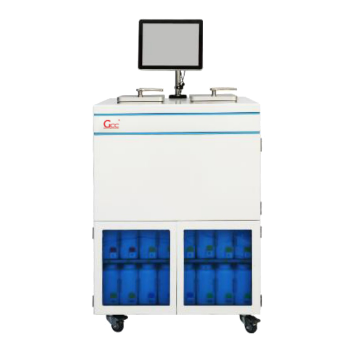
Phase 2: Tissue Processing and Dehydration
Tissue must undergo a series of chemical gradients to remove water and replace it with paraffin wax.
Automatic Tissue Processor: Modern laboratories favor enclosed vacuum processors to enhance dehydration efficiency and minimize reagent evaporation.
Key Features: High consistency, reagent protection, and user-friendly interface.
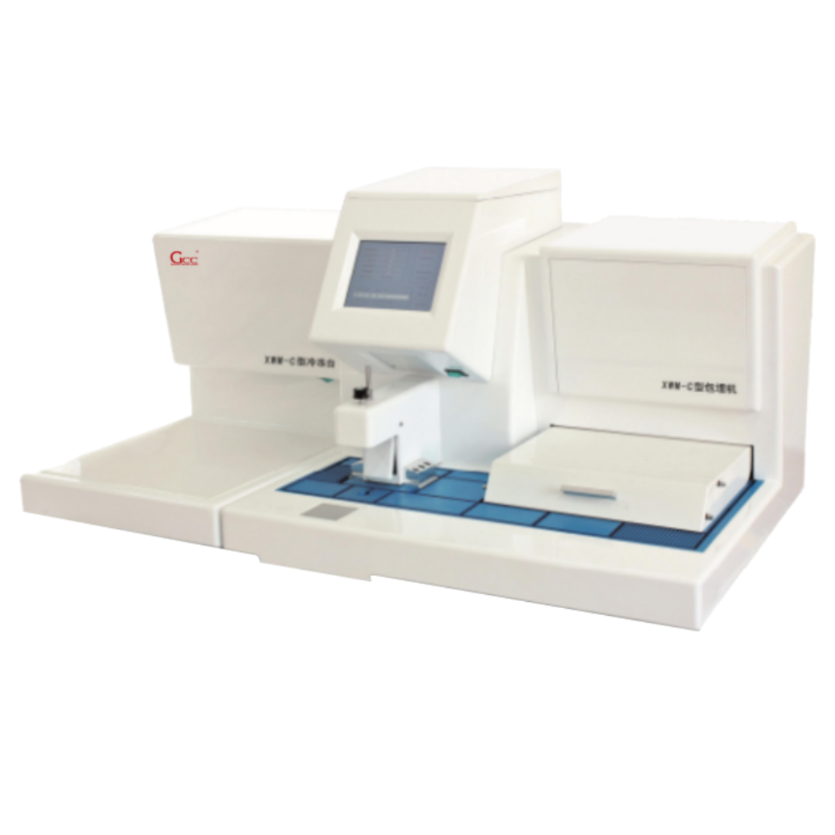
Phase 3: Tissue Embedding
Processed tissues are placed into metal molds, infused with molten paraffin, and cooled to form blocks that are easy to section.
Tissue Embedding Station: A complete embedding center typically includes a thermal console, a cryo console, and a paraffin dispenser.
Recommendation: GCC’s Modular Embedding Systems allow for flexible configurations to suit your lab space, ensuring smooth paraffin flow and precise temperature control.
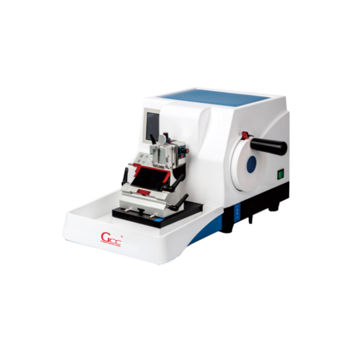
Phase 4: Precision Sectioning
This is the most technically demanding stage. The thickness of the section (typically 3-5 microns) directly determines the clarity under the microscope.
Microtome: Whether it is a rotary microtome for routine paraffin sections or a cryostat for rapid intraoperative diagnosis, mechanical stability is the top priority.
Water Bath & Slide Warmer: Used to flatten sections and adhere them securely to glass slides.
Phase 5: Staining and Mounting
The final step involves visualizing cellular structures through H&E staining or other special stains.
Automatic Stainer: Standardizes the staining process to eliminate color variations caused by manual operation.
Automated Coverslipper: Ensures long-term preservation of sections without air bubbles.
Laboratory Safety and Ventilation Systems
Throughout the entire laboratory setup, chemical safety is an essential background requirement. All chemical-intensive operations (such as staining and reagent preparation) should be conducted within professional Fume Hoods.
Why Choose GCC’s Integrated Solutions?
At GCC, we do more than just sell individual units; we provide Professional Integrated Engineering Solutions for pathology laboratories.
High-Standard Materials: All equipment is built using 304/316 grade stainless steel.
Custom-Engineered Fittings: We provide bespoke laboratory fittings tailored to the specific medical standards of different countries.
Global Delivery Expertise: With a strong presence in the Middle East and Southeast Asia, we possess mature experience in international logistics and on-site installation guidance.
Frequently Asked Questions
What equipment is needed for a hospital pathology lab?
Hospital pathology labs require grossing stations, tissue processors, embedding stations, microtomes, fume hoods, biosafety cabinets, staining systems, and microscopes based on their diagnostic scope. Hospital Pathology Lab Solutions
How to set up an independent pathology laboratory?
Setting up an independent lab requires proper facility planning, equipment selection, regulatory compliance, quality management systems, and qualified personnel. GCC Pathology offers turnkey solutions. Independent Lab Solutions
What are the different types of pathology grossing stations?
Grossing stations range from basic stainless steel workstations to advanced intelligent systems with touchscreen controls, formalin filling, and real-time environmental monitoring. The main types include basic models, height-adjustable stations, and smart digital systems. Types of Grossing Stations
�� Request Your Laboratory Configuration Plan
Planning a new pathology department or upgrading your existing equipment? GCC’s technical experts provide full-process support from planning to installation.
WhatsApp: +86 18148635992
Email: Sales@gzkunling.com
Website: www.gccpathology.com