坤灵最新logo-scaled-1.png)
Introduction
Microtomes are essential precision instruments in pathology laboratories, enabling the preparation of ultra-thin tissue sections for microscopic examination. From routine histopathology to advanced research applications, microtomes play a vital role in ensuring diagnostic accuracy.
This guide explores working principles, classifications, applications, and microtome maintenance best practices, helping laboratories improve performance, compliance, and efficiency.
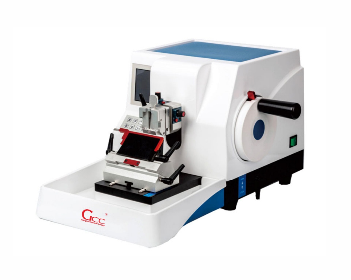
I. Definition and Overview of Microtomes
A microtome is a precision cutting instrument used to produce thin tissue sections (typically 1–50 μm) for microscopic analysis.
Key Terminology
- Microtome: Tissue sectioning instrument
- Sectioning: Cutting thin tissue slices
- Paraffin Embedding: Wax infiltration for support
- Cryosectioning: Frozen tissue cutting
- Ribbon: Continuous strip of tissue sections
II. Working Principle of Microtomes
Microtomes operate through controlled mechanical advancement + blade cutting:
- Sample Advancement: 1–50 μm per step
- Cutting Motion: Rotary / sliding / rocking
- Section Collection: Transfer to glass slides
- Temperature Control (Cryostat): -15°C to -30°C
III. Primary Functions in Pathology
- Diagnostic tissue preparation
- IHC and special staining
- Frozen section analysis
- Research and 3D reconstruction
- Biobanking and archival studies
IV. Structural Features and Materials
- Heavy-duty base (anti-vibration)
- Precision feed system
- Adjustable blade holder
- Safety lock & blade guard
- Ergonomic handwheel design
V. Classification and Technical Parameters
| Type | Thickness (μm) | Application | Power | Weight |
|---|---|---|---|---|
| Rotary | 1–60 | Routine histology | Manual / Motor | 25–40 kg |
| Cryostat | 5–50 | Frozen sections | 500–800W | 150–250 kg |
| Sliding | 1–100 | Large tissues | Manual | 30–50 kg |
| Ultramicrotome | 0.05–1 | Electron microscopy | 100–150W | 50–70 kg |
VI. Application Industries
- Hospitals & pathology labs
- Pharmaceutical companies
- Research institutes
- Veterinary labs
- Forensic laboratories
VII. Installation & Compliance
- Temperature: 18–22°C
- Humidity: 40–60% RH
- ISO 14644 cleanroom compliance
- GMP validation (IQ/OQ/PQ)
- IEC 61010 safety standards
VIII. Microtome Maintenance
Proper microtome maintenance is critical for ensuring section quality, instrument longevity, and regulatory compliance.
1. Daily Maintenance
- Clean debris after each use
- Wipe surfaces with 70% ethanol
- Remove paraffin residues
- Check blade condition
2. Weekly Maintenance
- Inspect blade holder alignment
- Clean feed mechanism
- Lubricate moving components (if required)
3. Monthly Maintenance
- Verify section thickness accuracy
- Inspect mechanical stability
- Check handwheel locking system
4. Annual Maintenance
- Full calibration by certified technician
- Replace worn components
- Verify safety systems
5. Cryostat-Specific Maintenance
- Defrost regularly
- Replace HEPA filters (every 3–6 months)
- Clean internal chamber to prevent contamination
Microtome Maintenance Best Practices
- Use only manufacturer-approved consumables
- Keep detailed maintenance logs (GMP requirement)
- Train operators for proper handling
- Schedule preventive maintenance
👉 Key Insight:
Proper maintenance can reduce downtime by 30–40% and significantly improve section consistency.
IX. Which Microtome Is Right for Your Lab? A Selection Guide by Lab Type
Selecting the wrong microtome type is one of the most common and costly mistakes when setting up a pathology laboratory. Different laboratory environments have distinct workflow requirements, specimen volumes, and diagnostic responsibilities. Choosing the appropriate microtome ensures optimal section quality, operational efficiency, and long-term return on investment.
Recommended Microtome Types by Laboratory
| Laboratory Type | Recommended Microtome | Why It Fits |
|---|---|---|
| Hospital Pathology Department | Fully Automatic Rotary Microtome | High throughput, excellent section consistency, ideal for routine paraffin-embedded tissue processing |
| University Teaching Hospital | Semi-Automatic Rotary Microtome + Cryostat | Supports both resident training and clinical diagnostics while balancing automation and hands-on learning |
| Intraoperative Frozen Section Laboratory | Cryostat Microtome | Produces frozen sections within 10–15 minutes, enabling rapid surgical decision-making |
| Research Laboratory | Vibrating Microtome (Vibratome) | Allows sectioning of fresh or living tissues without paraffin embedding, preserving cellular structures |
| Small Clinics & Veterinary Laboratories | Desktop Semi-Automatic Microtome | Compact footprint, cost-effective operation, suitable for moderate specimen volumes |
Choosing Between Rotary and Cryostat Microtomes
A rotary microtome handles the vast majority of routine histopathology applications efficiently and remains the most widely used microtome type worldwide. However, laboratories performing intraoperative consultations require a dedicated cryostat system because frozen sectioning and paraffin sectioning are fundamentally different processes and cannot be performed using the same instrument.
For university pathology departments and teaching hospitals that combine clinical workloads with resident education, a semi-automatic rotary microtome offers a practical balance. Operators can learn essential sectioning techniques while benefiting from automation that improves productivity and reproducibility.
Budget Considerations and Equipment Planning
Budget planning is another important consideration. Clinical-grade rotary microtomes typically range from USD 8,000–22,000, while high-performance cryostats designed for ISO 15189 and modern pathology environments generally range from USD 15,000–35,000, depending on temperature stability, automation level, and safety features.
If you are unsure which microtome configuration best suits your specimen volume, staffing level, or diagnostic workflow, GCC Pathology can provide a customized recommendation based on your laboratory’s weekly case load and operational requirements.
Request Microtome Specifications & Pricing for Your Lab
Whether you are establishing a new pathology laboratory, upgrading existing equipment, or expanding frozen section capabilities, GCC Pathology can help you identify the most suitable microtome solution.
Our Support Includes
- Rotary Microtome Selection & Comparison
- Cryostat Configuration Recommendations
- Histopathology Workflow Optimization
- GMP & ISO-Compliant Laboratory Equipment
- Installation, Validation, and Training Support
→ Request Microtome Specifications & Pricing for Your Lab
→ Compare Rotary vs Cryostat Models
Contact GCC Pathology
WhatsApp: https://wa.me/8618148635992
Email: Victor@gccpathology.com
Our specialists will recommend the most suitable microtome based on your laboratory’s specimen volume, workflow requirements, and budget.
Frequently Asked Questions
How often should pathology equipment be serviced?
Most pathology equipment requires annual professional servicing, with daily operator checks and monthly preventive maintenance. High-use equipment like microtomes and tissue processors may need more frequent attention. Equipment Maintenance Guide
When is it time to replace vs repair pathology equipment?
Consider replacement when repair costs exceed 50% of replacement value, when parts are no longer available, or when newer technology offers significant workflow or safety improvements. Repair vs Replace Guide
What is a microtome and how does it work?
A microtome is a precision instrument used to cut extremely thin tissue sections (typically 2-10 micrometers) for microscopic examination. It advances the specimen block across a blade with micron-level accuracy. Microtome Guide
What are the different types of pathology grossing stations?
Grossing stations range from basic stainless steel workstations to advanced intelligent systems with touchscreen controls, formalin filling, and real-time environmental monitoring. The main types include basic models, height-adjustable stations, and smart digital systems. Types of Grossing Stations
How does a tissue processor work in histopathology?
A tissue processor automates the dehydration, clearing, and paraffin infiltration of tissue specimens. It moves samples through graded alcohols, xylene, and molten paraffin under controlled temperature and vacuum conditions. What is a Tissue Processor