坤灵最新logo-scaled-1.png)
Abstract: This article provides a comprehensive technical overview of tissue processors, essential instruments in modern histopathology laboratories. It covers the working principles, functionalities, structural characteristics, application scenarios, advantages, and maintenance protocols of these critical devices, with emphasis on their role in diagnostic precision and laboratory efficiency.
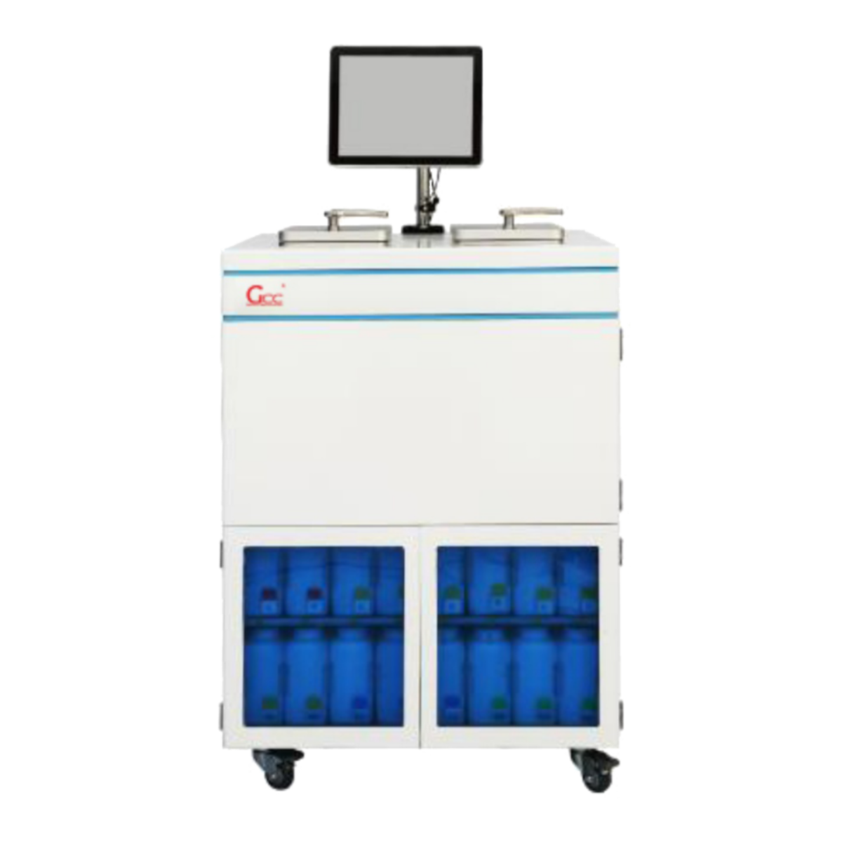
Definition and Overview
A tissue processor is an automated instrument designed for the preparation of biological tissue specimens for microscopic examination through histopathological analysis. This sophisticated apparatus automates the multi-step process of tissue fixation, dehydration, clearing, and infiltration with paraffin wax, which is essential for producing high-quality tissue sections for diagnosis.
Working Principle
Tissue processors operate on the principle of sequential chemical processing through either mechanical transfer or fluid transfer methodologies:
- Mechanical Transfer Systems: Tissue cassettes are moved through stationary reagent containers via a robotic arm or carousel mechanism.
- Fluid Transfer Systems: Tissues remain stationary while reagents and paraffin are pumped into and out of a single processing chamber.
The processing cycle typically follows these stages: fixation in formalin, dehydration through graded alcohols (ethanol or isopropanol), clearing with xylene or xylene substitutes, and infiltration with molten paraffin wax. Modern processors utilize microprocessors to control temperature, pressure, and vacuum cycles to optimize processing efficiency and quality.
Main Functions and Roles
Tissue processors serve several critical functions in histopathology laboratories:
- Automation of tissue processing, ensuring consistency and reproducibility
- Standardization of processing protocols across specimens and operators
- Enhancement of laboratory efficiency through simultaneous processing of multiple specimens
- Reduction of technician exposure to hazardous chemicals
- Improvement of tissue morphology preservation for accurate diagnosis
- Enablement of specialized processing protocols for specific tissue types
Structural Characteristics and Materials
Modern tissue processors feature robust construction with materials selected for chemical resistance and durability:
- Processing Chamber: Constructed from stainless steel or chemical-resistant polymers
- Reagent Containers: Made from polypropylene, polycarbonate, or glass with chemical resistance
- Fluid Path Components: Utilizing PTFE, Viton, or other chemically inert materials
- Heating Systems
- Sealing Systems: Hermetic seals to contain fumes and maintain vacuum/pressure
- User Interface: Touchscreen displays with programmable protocols and data logging
Classification and Technical Parameters
| Parameter | Standard Models | High-Capacity Models | Compact Models |
| Cassette Capacity | 150-300 | 300-600 | 50-150 |
| Power Consumption | 1200-1500W | 1800-2200W | 800-1000W |
| Filtration Level | HEPA H13 | HEPA H14 | HEPA H13 |
| Dimensions (W×D×H, cm) | 80×70×110 | 100×80×130 | 60×50×90 |
| Airflow Rate (m³/h) | 150-200 | 250-350 | 100-150 |
| Reagent Stations | 12-16 | 16-24 | 8-12 |
Typical Application Industries
Tissue processors are primarily employed in:
- Hospital histopathology departments
- Reference and diagnostic laboratories
- Academic and research institutions
- Veterinary pathology laboratories
- Pharmaceutical research and development
- Forensic pathology facilities
Installation and Usage Considerations
Proper installation and operation are critical for optimal performance and compliance with international standards:
- Installation must comply with GMP (Good Manufacturing Practice) and ISO 14644 cleanroom standards where applicable
- Adequate ventilation with fume extraction systems meeting OSHA requirements
- Leveled installation on vibration-resistant surfaces
- Access to appropriate power sources with emergency power backup for cycle completion
- Environmental controls maintaining temperature at 18-22°C and humidity below 60%
- Validation protocols following installation (IQ/OQ/PQ)
- Regular calibration of temperature sensors and timers
- Comprehensive staff training on operation and safety procedures
Maintenance and (Maintenance and Care Recommendations)
A structured maintenance program is essential for reliable operation:
- Daily: Visual inspection for leaks, verification of reagent levels, and surface disinfection
- Weekly: Cleaning of wax reservoirs, checking of tubing integrity, and verification of vacuum/pressure systems
- Monthly: Deep cleaning of processing chambers, calibration of temperature sensors, and testing of safety interlocks
- Quarterly: Replacement of air filters, comprehensive system diagnostics, and seal inspections
- Biannually: Replacement of fluid transfer tubing, pump maintenance, and electrical safety testing
- Annually: Full system calibration, preventive parts replacement, and validation per manufacturer specifications
- As needed: Replacement of UV lamps in biocontainment models and updating of software systems
All maintenance activities should be documented in compliance with quality management systems, and any malfunction should be addressed immediately to prevent compromise of tissue specimens.
Frequently Asked Questions
How often should pathology equipment be serviced?
Most pathology equipment requires annual professional servicing, with daily operator checks and monthly preventive maintenance. High-use equipment like microtomes and tissue processors may need more frequent attention. Equipment Maintenance Guide
When is it time to replace vs repair pathology equipment?
Consider replacement when repair costs exceed 50% of replacement value, when parts are no longer available, or when newer technology offers significant workflow or safety improvements. Repair vs Replace Guide
How does a tissue processor work in histopathology?
A tissue processor automates the dehydration, clearing, and paraffin infiltration of tissue specimens. It moves samples through graded alcohols, xylene, and molten paraffin under controlled temperature and vacuum conditions. What is a Tissue Processor
What are the different types of pathology grossing stations?
Grossing stations range from basic stainless steel workstations to advanced intelligent systems with touchscreen controls, formalin filling, and real-time environmental monitoring. The main types include basic models, height-adjustable stations, and smart digital systems. Types of Grossing Stations
What is a microtome and how does it work?
A microtome is a precision instrument used to cut extremely thin tissue sections (typically 2-10 micrometers) for microscopic examination. It advances the specimen block across a blade with micron-level accuracy. Microtome Guide